SkinCheck for Health Insurance Instantly
- SkinCheck is your skin health systemised to put you at ease.
- We offer great service and reassurance in our welcoming clinic centrally located at Østerport Station by Dr Inanna Weiss, one of Denmark’s most experienced skin cancer specialists.
Years of experience have taught us that early detection of skin cancer is important. That’s why we offer an easily accessible full body examination with mapping of all skin lesions to increase the chances of early detection of skin cancer and melanoma.
Call us on 61 10 18 10 or book online
This is SkinCheck:
How does the examination work?
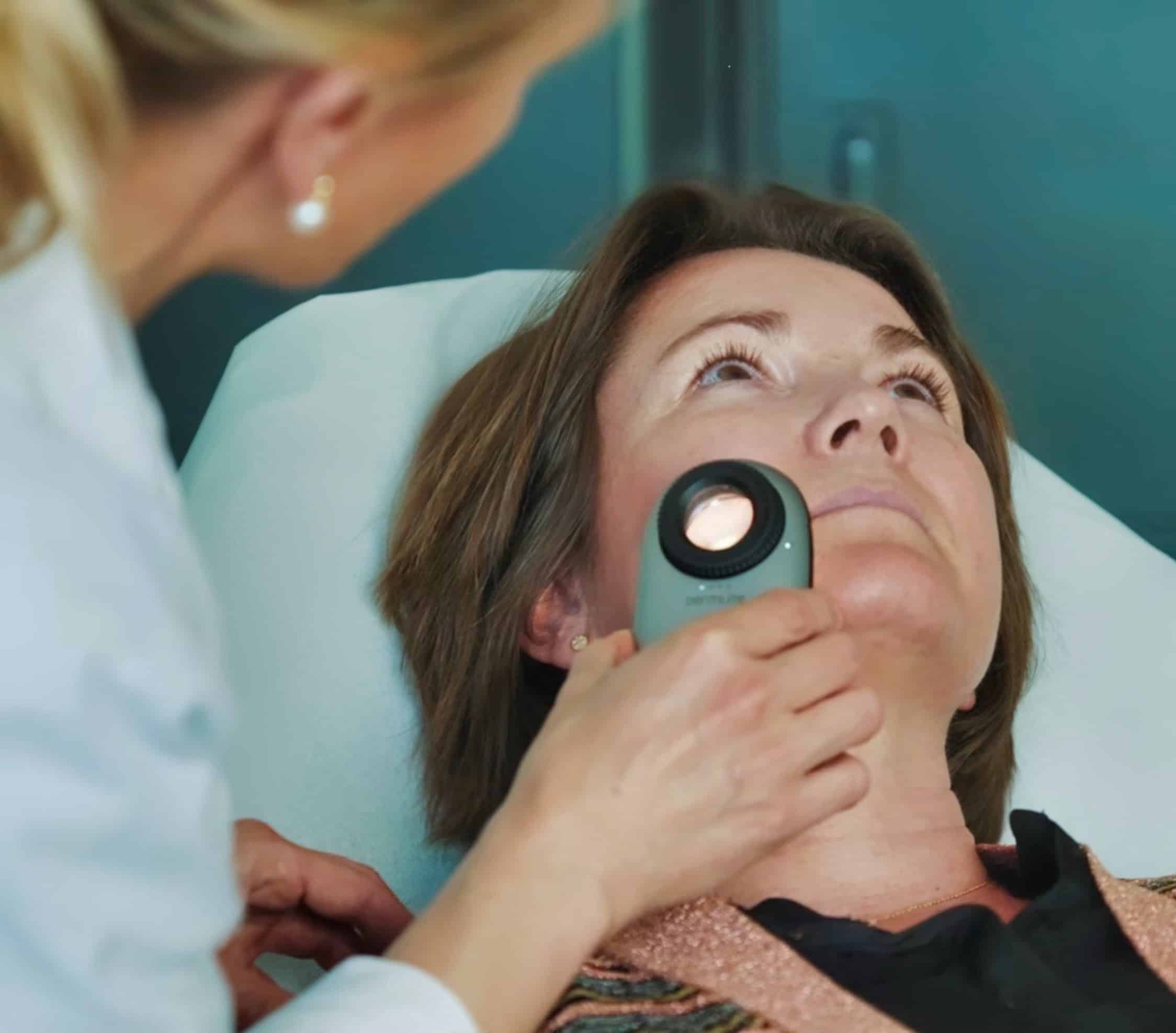
The examination takes between 25-45 minutes and is performed by a skin cancer specialist. You will be asked to strip down to your underwear. You will then be examined from head to toe with a dermatoscope.
Any moles, spots or lumps will be assessed using the dermatoscope – a medical instrument that illuminates and magnifies your skin. The light is polarised so that it can penetrate the top layer of skin more easily, allowing the doctor to see features that would otherwise not be visible, enabling the detection of subtle signs of skin cancer.
You will then be photographed for mole mapping: this is whole body photography that is supplemented with single dermoscopic images of individual skin lesions and is an advanced method of monitoring moles (nevi) and other skin lesions over time. In particular, this technique is used to identify changes that may indicate skin cancer, including melanoma, at an early stage.
Mole mapping:
The whole body or specific areas of the skin are photographed with a specialised camera. This is complemented by digital dermoscopy, where dermoscopic images are taken of selected skin lesions. The images are uploaded to a specialised software that helps analyse and track the development of the moles over time. The images form the basis of a baseline against which future images can be compared. The software can compare new images with baseline images and identify changes in shape, size, colour and structure. At the next visit when new images are taken, the software will detect changes in existing skin changes and detect new skin changes.


